Purpose
Using various imaging modalities, including Adaptive Optics Scanning Laser Ophthalmoscope (AOSLO) and OCT-Angiography (OCTA), we aim to understand blood vessel response in diabetic patients with ischemic retinal diseases such as diabetic retinopathy.
OCT-A Imaging
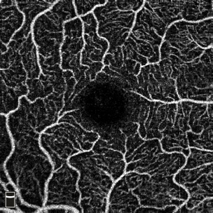
OCT-Angiography is a non-invasive technique used to image blood vessels in the superficial, middle, and deep capillary plexuses. Using this modality, we are able to obtain a blood vessel map of the macula while obtaining information on blood flow throughout the retina. Dr. Fawzi’s team currently uses OCT-Angiography to learn more about diabetic retinopathy and dark adaptation.
AOSLO Imaging

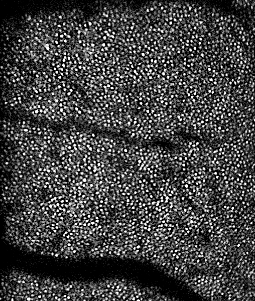
Adaptive Optics Scanning Laser Ophthalmoscopy (AOSLO) is the newest, non-invasive imaging technique used to visualize the retina. AOSLO functions much like a microscope, but uses the light reflected off the retina to capture high-resolution images in-vivo. Each layer of the retina can be imaged individually, which allows us to visualize not only the blood vessels in the superficial layer, but the rods and cones in the photoreceptor layer. Dr. Fawzi’s team uses AOSLO to investigate how blood flow changes in diabetic patients.